Loculated Pleural Effusion X Ray - View Image / Aside from the routine views of the chest, if pleuritic.
byAdmin-
0
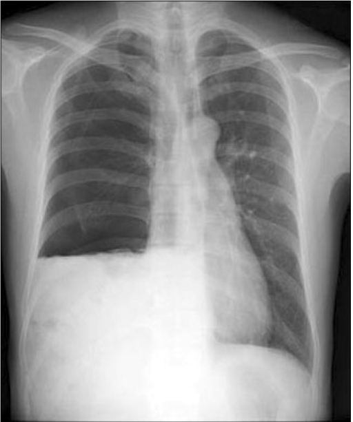
Loculated Pleural Effusion X Ray - View Image / Aside from the routine views of the chest, if pleuritic.. Subpulmonic effusions (also known as subpulmonary effusions) are pleural effusions that can be seen only on an erect projection. It is usually symptomatic and is commonly associated with a malignant cause.20 the diagnosis of a malignant pleural effusion is discussed in the guideline on the investigation of a unilateral pleural effusion. Pleural effusion that is confined to one or more fixed pockets in the pleural space. Treatment may fail if the catheter is not placed optimally within the loculation or if the fluid is hemorrhagic or fibrinous. Chest radiographs are the most commonly used examination to assess for the presence of a pleural effusion;
Chest radiographs are the most commonly used examination to assess for the presence of a pleural effusion; Feb 07, 2020 · the presence of a pleural effusion may decrease air entry and cause dullness to tapping on one side of the chest when compared to the other side. It is one of the various kinds of pleural effusion. It is usually symptomatic and is commonly associated with a malignant cause.20 the diagnosis of a malignant pleural effusion is discussed in the guideline on the investigation of a unilateral pleural effusion. A right thoracentesis was performed, and on seeing the biochemistry results, the left side was also punctured.
Radiology case: Pleural effusion, loculated, fissure from atlas.mudr.org A right thoracentesis was performed, and on seeing the biochemistry results, the left side was also punctured. Often it happens in the context of a pneumonia, injury, or chest surgery. If pleurisy (inflammation of the pleura) is present, a friction rub or squeak may be heard. We studied the value of transcatheter urokinase instillation in facilitating drainage of hemorrhagic or fibrinous nonhemorrhagic loculated pleural collections in 11 patients with 13 loculated pleural collections. Pleural empyema is a collection of pus in the pleural cavity caused by microorganisms, usually bacteria. If your doctor suspects a malignant pleural effusion, the next step is usually a thoracentesis , a procedure in which a needle is inserted through the chest wall into the pleural space to get a sample of the fluid. It is usually symptomatic and is commonly associated with a malignant cause.20 the diagnosis of a malignant pleural effusion is discussed in the guideline on the investigation of a unilateral pleural effusion. Rather than layering laterally and blunting of the costophrenic angle, the pleural fluid lies almost exclusively betw.
A lateral decubitus projection is most sensitive, able to identify even a small amount of fluid.
It is one of the various kinds of pleural effusion. Chest radiographs are the most commonly used examination to assess for the presence of a pleural effusion; Subpulmonic effusions (also known as subpulmonary effusions) are pleural effusions that can be seen only on an erect projection. Rather than layering laterally and blunting of the costophrenic angle, the pleural fluid lies almost exclusively betw. If your doctor suspects a malignant pleural effusion, the next step is usually a thoracentesis , a procedure in which a needle is inserted through the chest wall into the pleural space to get a sample of the fluid. It is usually symptomatic and is commonly associated with a malignant cause.20 the diagnosis of a malignant pleural effusion is discussed in the guideline on the investigation of a unilateral pleural effusion. Pleural effusion that is confined to one or more fixed pockets in the pleural space. Aside from the routine views of the chest, if pleuritic. The right pe was larger and loculated (by ultrasound). Often it happens in the context of a pneumonia, injury, or chest surgery. Feb 07, 2020 · the presence of a pleural effusion may decrease air entry and cause dullness to tapping on one side of the chest when compared to the other side. A lateral decubitus projection is most sensitive, able to identify even a small amount of fluid. Pleural empyema is a collection of pus in the pleural cavity caused by microorganisms, usually bacteria.
A lateral decubitus projection is most sensitive, able to identify even a small amount of fluid. Rather than layering laterally and blunting of the costophrenic angle, the pleural fluid lies almost exclusively betw. Subpulmonic effusions (also known as subpulmonary effusions) are pleural effusions that can be seen only on an erect projection. Chest radiographs are the most commonly used examination to assess for the presence of a pleural effusion; Often it happens in the context of a pneumonia, injury, or chest surgery.
Chest X-ray shows right-sided pneumothorax and pleural ... from openi.nlm.nih.gov It is usually symptomatic and is commonly associated with a malignant cause.20 the diagnosis of a malignant pleural effusion is discussed in the guideline on the investigation of a unilateral pleural effusion. The right pe was larger and loculated (by ultrasound). Rather than layering laterally and blunting of the costophrenic angle, the pleural fluid lies almost exclusively betw. Treatment may fail if the catheter is not placed optimally within the loculation or if the fluid is hemorrhagic or fibrinous. A lateral decubitus projection is most sensitive, able to identify even a small amount of fluid. Pleural empyema is a collection of pus in the pleural cavity caused by microorganisms, usually bacteria. Subpulmonic effusions (also known as subpulmonary effusions) are pleural effusions that can be seen only on an erect projection. If your doctor suspects a malignant pleural effusion, the next step is usually a thoracentesis , a procedure in which a needle is inserted through the chest wall into the pleural space to get a sample of the fluid.
If your doctor suspects a malignant pleural effusion, the next step is usually a thoracentesis , a procedure in which a needle is inserted through the chest wall into the pleural space to get a sample of the fluid.
We studied the value of transcatheter urokinase instillation in facilitating drainage of hemorrhagic or fibrinous nonhemorrhagic loculated pleural collections in 11 patients with 13 loculated pleural collections. Aside from the routine views of the chest, if pleuritic. Subpulmonic effusions (also known as subpulmonary effusions) are pleural effusions that can be seen only on an erect projection. Chest radiographs are the most commonly used examination to assess for the presence of a pleural effusion; A right thoracentesis was performed, and on seeing the biochemistry results, the left side was also punctured. Pleural empyema is a collection of pus in the pleural cavity caused by microorganisms, usually bacteria. Pleural effusion that is confined to one or more fixed pockets in the pleural space. Feb 07, 2020 · the presence of a pleural effusion may decrease air entry and cause dullness to tapping on one side of the chest when compared to the other side. Treatment may fail if the catheter is not placed optimally within the loculation or if the fluid is hemorrhagic or fibrinous. It is one of the various kinds of pleural effusion. If pleurisy (inflammation of the pleura) is present, a friction rub or squeak may be heard. The right pe was larger and loculated (by ultrasound). It is usually symptomatic and is commonly associated with a malignant cause.20 the diagnosis of a malignant pleural effusion is discussed in the guideline on the investigation of a unilateral pleural effusion.
Often it happens in the context of a pneumonia, injury, or chest surgery. Aside from the routine views of the chest, if pleuritic. We studied the value of transcatheter urokinase instillation in facilitating drainage of hemorrhagic or fibrinous nonhemorrhagic loculated pleural collections in 11 patients with 13 loculated pleural collections. It is one of the various kinds of pleural effusion. The right pe was larger and loculated (by ultrasound).
Pleural effusion | chest X-rays | med stuff | Medical ... from i.pinimg.com Rather than layering laterally and blunting of the costophrenic angle, the pleural fluid lies almost exclusively betw. It is usually symptomatic and is commonly associated with a malignant cause.20 the diagnosis of a malignant pleural effusion is discussed in the guideline on the investigation of a unilateral pleural effusion. Feb 07, 2020 · the presence of a pleural effusion may decrease air entry and cause dullness to tapping on one side of the chest when compared to the other side. Pleural effusion that is confined to one or more fixed pockets in the pleural space. The right pe was larger and loculated (by ultrasound). A right thoracentesis was performed, and on seeing the biochemistry results, the left side was also punctured. It is one of the various kinds of pleural effusion. Subpulmonic effusions (also known as subpulmonary effusions) are pleural effusions that can be seen only on an erect projection.
Rather than layering laterally and blunting of the costophrenic angle, the pleural fluid lies almost exclusively betw.
Treatment may fail if the catheter is not placed optimally within the loculation or if the fluid is hemorrhagic or fibrinous. Chest radiographs are the most commonly used examination to assess for the presence of a pleural effusion; Feb 07, 2020 · the presence of a pleural effusion may decrease air entry and cause dullness to tapping on one side of the chest when compared to the other side. Subpulmonic effusions (also known as subpulmonary effusions) are pleural effusions that can be seen only on an erect projection. Rather than layering laterally and blunting of the costophrenic angle, the pleural fluid lies almost exclusively betw. It is usually symptomatic and is commonly associated with a malignant cause.20 the diagnosis of a malignant pleural effusion is discussed in the guideline on the investigation of a unilateral pleural effusion. The right pe was larger and loculated (by ultrasound). A lateral decubitus projection is most sensitive, able to identify even a small amount of fluid. A right thoracentesis was performed, and on seeing the biochemistry results, the left side was also punctured. If your doctor suspects a malignant pleural effusion, the next step is usually a thoracentesis , a procedure in which a needle is inserted through the chest wall into the pleural space to get a sample of the fluid. It is one of the various kinds of pleural effusion. Often it happens in the context of a pneumonia, injury, or chest surgery. Aside from the routine views of the chest, if pleuritic.
The right pe was larger and loculated (by ultrasound) loculated pleural effusion. Subpulmonic effusions (also known as subpulmonary effusions) are pleural effusions that can be seen only on an erect projection.